Case Challenge: An Increasing Problem in Melbourne
Case Challenge: An Increasing Problem in Melbourne
Dr Robyn Troutbeck
A 35 year old female presents with a 6 day history of photopsias and a central scotoma in the LE. Her best corrected vision was R 6/6 L 6/120. Her anterior segments were quiet and she had 1+ ant vit cell in both eyes.
The colour photos presented below did not show any obvious fundus abnormalities.
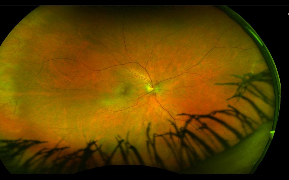
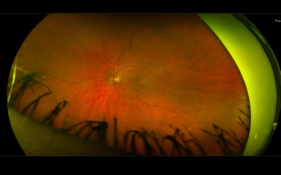
A very useful modality is fundus autofluorescence to examine the outer retina and RPE. We need to explain why her vision is so poor in the LE.
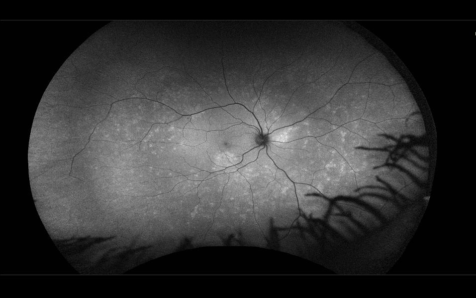
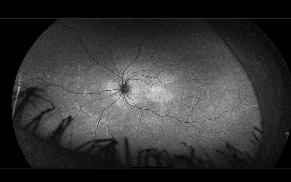
These photos show extensive changes in both eyes but more prominent at the L macula which accounts for the decreased vision.
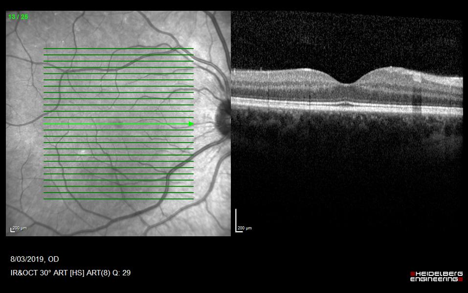
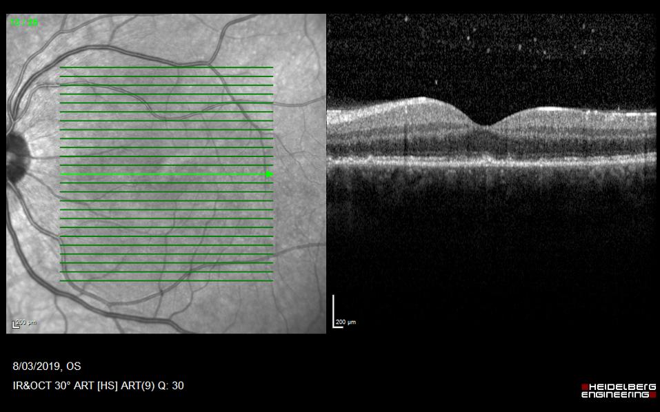
OCT confirms outer retinal changes at the level of the ellipsoid zone in the L macula.
Further history from the patient revealed a 3 month history of a non-healing mouth ulcer with associated submandibular lymphadenopathy. Bloods gave us the answer – syphilis serology was positive. Fortunately after 2 weeks of intravenous penicillin her vision improved. The incidence of syphilis is increasing rapidly in Victoria and all patients with uveitis involving the posterior segment need to be screened.