Elderly Man with Subtle Macular Changes
Case Study: Elderly Man with Subtle Macular Changes
Dr Daniel Chiu 
Presentation
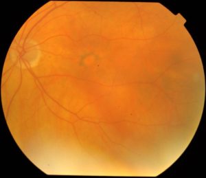
70yo male with LVA 6/12 on asymptomatic eye check. RVA is normal with subtle macular pigment disturbance only
What do you see in the fundus photo?
1. Macular naevus
2. Macular hole
3. Macular RPE pigment clumping
4. Optic atrophy
5. Inferior retinal detachment
A. 1 & 4
B. 2 & 5
C. 2 alone
D. 2 & 4
E. 3 alone
Click for answer
Answer E.
(None of the features except 3 is really identifiable in the colour photo)
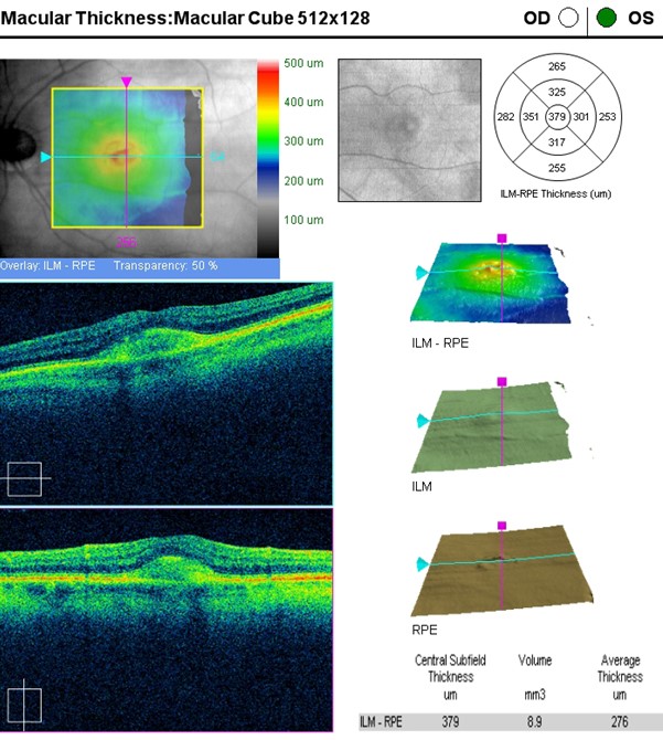
What do you see in this macular OCT?

1. Macular schisis
2. Choroidal neovascular membrane
3. Subfoveal hyperreflective deposit
4. Subfoveal fibrous scar with intraretinal fluid
5. Temporal choroidal naevus with retinal thinning on the map
6. Macular oedema
A. 1 & 5
B. 3 alone
C. 2 alone
D. 4 & 6
E. 5 alone
Click for answer
Answer B.
(None of the features 1-6 except 3 is really identifiable in the OCT. Central retinal thickness excluding the dome-shape smooth subfoveal deposit is really quite normal. Machine interpreted figure of 379um central subfield thickness if not a real representation of the foveal retinal tissue thickness)
What is the likely diagnosis in this case?
1. Macular choroidal polyps
2. Submacular drusenoid pigment epithelial detachment
3. Active submacular choroidal neovascularization
4. Adult vitelliform macular dystrophy
5. North Carolina macular dystrophy
Click for answer
Answer 4.
(The fundus photo and the OCT is classic picture or adult (or acquired) vitelliform macular dystrophy. Very common to have “pigment figures” surround the circular or ovoid subfoveal vitelliform deposit. This condition is very often mistaken as neovascular AMD in elderly patient and wrongly treated with months and months of anti-VEGF with no real resolution or worsening!!)