Elderly Man with Gradual Visual Change
Case Study: Elderly Man with Gradual Visual Change.
Dr Daniel Chiu 
Presentation
60 yo male with gradual change in central vision becoming more rapid in the last 2 weeks.
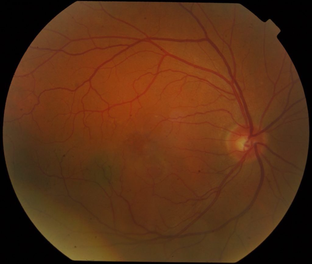
What is showing in the colour photo?
1. Large soft drusen
2. Inferotemporal retinal detachment
3. Parafoveal pigment epithelial detachment
4. Inferotemporal retinoschisis
5. Paramacular choroidal naevus
A. 1 & 2
B. 3 & 5
C. 1 & 4
D. 3 & 5
E. 1, 2, 3, 4 & 5
Click for answer
Answer D.
(Only very few small drusens in upper temporal macula which are not classified as soft drusens. Inferotemporal light shadow is photographic artifact, not detachment or schisis. Temporal macular darkened area is consistent with naevus and faint circular area with clear centre below fovea is classic appearance of pigment epithelial detachment)
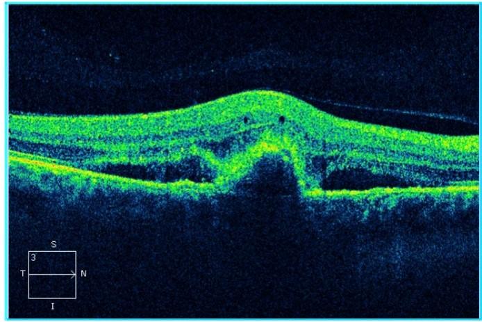
What is showing in this macular OCT?
1. Subretinal haemorrhage
2. Subretinal heperreflective material (SHRM)
3. Pigment epithelial detachment
4. Choroidal neovascular membrane
5. Intraretinal cyst
6. Posterior vitreous detachment
7. Subretinal fluid
A. 1 & 5
B. 2 & 4
C. 5, 6 & 7
D. 3 & 4
E. 2, 3, 5 & 7
F. 1, 2, 3, 4, 5, 6 & 7
Click for answer
E.
(SHRM is the descriptive terms for the dense reflective substance in the outer retina, likely representing exudative material from CNVM. In this picture, is is capping the dome shape pigment epithelial detachment. One cannot diagnose subretinal haemorrhage from OCT alone. There is a line of posterior hyaloid elevated from the nasal retinal surface but it does not mean there is PVD. Again diagnosis of CNVM can only be inferred from the OCT, but CNVM is not directly shown as such on OCT. Subretinal fluid around the PED is self evident in the picture.)
What is / are the diagnosis / differential diagnoses?
1. central serous chorioretinopathy
2. Occult choroidal neovascularisation
3. choroidal melanoma
4. polypoidal vasculopathy
5. All of the above
A. 1 & 2
B. 1 & 4
C. 3 alone
D. 4 alone
E. 2 & 4
F. 5 alone
Click for answer
E.
(SHRM is very seen in CSC, naevus is very small and appear very flat so melanoma is unlikely and OCT does not support that. Occult CNV cannot be excluded and choroidal polyps can share all these features and should be considered especially if AMD features such as soft drusens are not prominent.)