A PICture Tells a Thousand Words
Case Study:
Dr Robyn Troutbeck
Presentation
A 23 year old female presents with sudden distortion in her left eye. She is a low myope and has never had any eye problems in the past. Her vision is R and L 6/5. Her R fundus is normal and the L has a small central scar with a small haemorrhage.
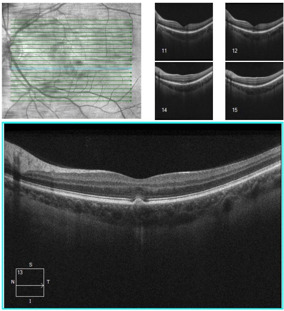
Her OCT is shown below
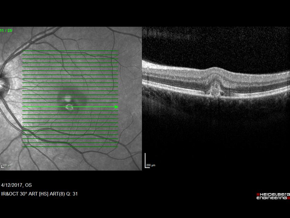
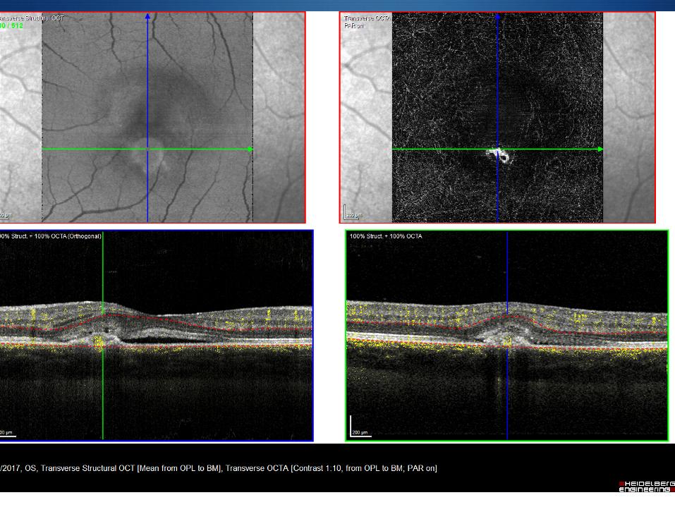
The OCT shows subretinal fluid under the macula and an inferior area of SHRM (subretinal hyper reflective material). This is consistent with a CNV likely secondary to a PIC lesion. PIC or punctate inner choroidopathy is not uncommon in myopic females and is usually asymptomatic unless a CNV develops, Rather than performing an angiogram to confirm the diagnosis she had an OCTA.
What treatment could we offer her?
Click for answer.
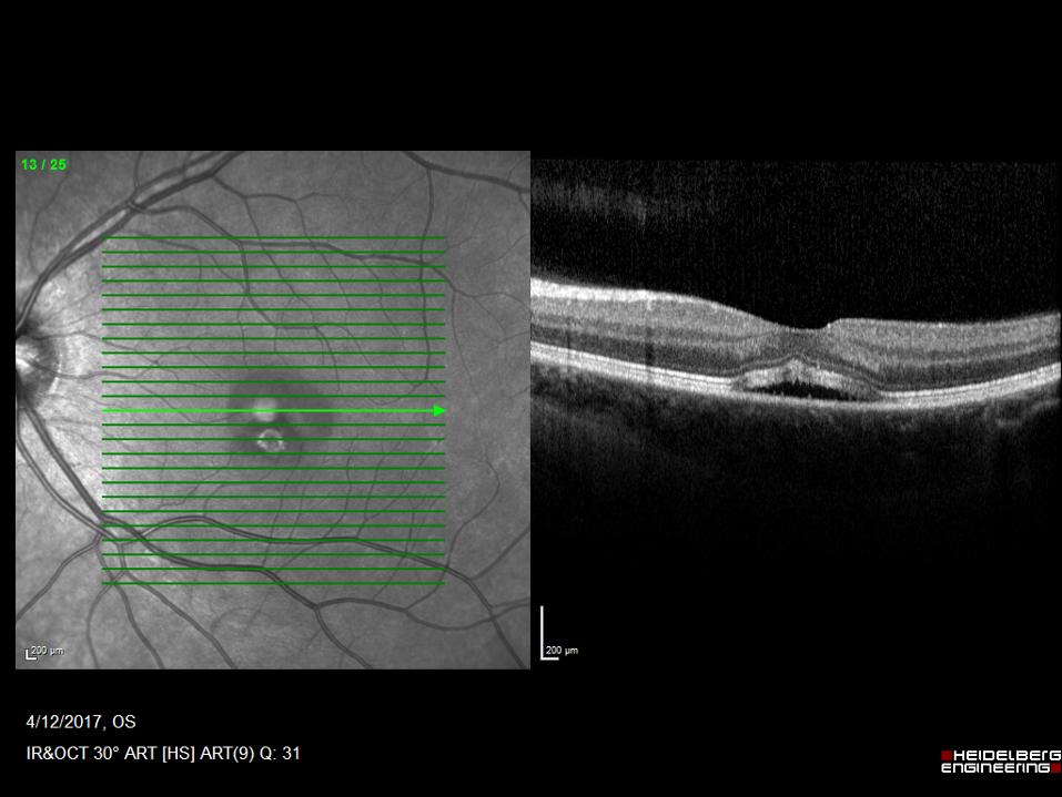
The best treatment is an anti-VEGF. The scarring looked old and there were no signs of active inflammation. We had an open discussion about the risks of anti-VEGF in females of child bearing age and we confirmed that she was not pregnant. She had an injection and 4 weeks later was reviewed. Her OCT is now is dry and with resolution of the fluid, her distortion has resolved. She may require further treatment in the future, but unlike AMD does not need regular treatment. Her post injection OCT is shown below.