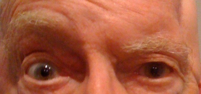
Droopy Eyelid
Case Study:
Dr Charles Su
Presentation
This 76 year old male presented with a complaint of a droopy left upper lid.
Indeed, there is skin hooding over the lashes on that side.

What 4 other relevant features do you see which show that this is not likely to be simply involutional upper lid skin excess ?
Click for answer
1. Marked asymmetry. A unilateral process is much less likely to be caused by involutional changes.
2. Left Brow ptosis. This shows that more than the upper lid is involved. In fact, the brow ptosis is the reason for the skin hooding.
3. Loss of forehead creases on the left. This indicates reduced contraction of the left frontalis muscle. This explains the left relative brow ptosis. The right side is unaffected.
4. A depression in the temporal scalp area lateral to the brow on the left. This might be a site of previous surgery, or trauma, or it might be deformed by some other process, such as an infiltrative process. This region is traversed by the frontal branch of the facial nerve, which lies superficial to the deep temporal fascia covering the temporalis muscle.
What process are we dealing with ?
Click for answer
We are dealing with a Left Paretic Brow Ptosis. A search must be made for a cause of the facial paresis, if one is not evident from the patient’s history. Even if the patient had surgery in that region, if the onset of the paresis appears to have come later, then surgical trauma cannot be blamed for it. For example, if the surgery was done to excise a malignancy such as a squamous cell carcinoma (SCC), a later nerve paresis there might indicate a recurrence of the carcinoma infiltrating the local nerve. SCCs are known to exhibit perineural infiltration, and hence any motor or sensory deficit which has no other convincing explanation needs to be investigated. An MRI scan can detect thickening of the affected nerve, and sometimes a local biopsy or a biopsy of the nerve involved is required to make the diagnosis, where the suspicion of such a process warrants it.