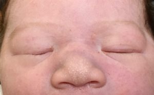
Progressively Enlarging Lid Lump in a Baby
Case Study:
Dr Elaine Wong
Presentation
8 week old baby present with a right medial lid lump that is progressively larger and becoming more noticeable. Mother has not noticed any redness in the surrounding area and denies any injuries. The lesion did not seem to bother the baby either.
On examination, the baby was able to maintain central and steady gaze with either eye. There are no signs of hypoglobus or strabismus. There is no mass effect from the lesion.
On palpation, there is a 5mm x 5mm soft lump superonasally attached to the orbital rim. There is no erythema or swelling in the surround skin.

Newborn.

8 weeks old.
What is this?
Click here for answer.
The location and presentation are most consistent of a dermoid cyst.
What are the differential diagnosis?
Click here for answer.
Mucocoele or encephalocoele
Discussion
Dermoid cyst is the most common space-occupying orbital lesion in the childhood. It is a result of sequestration of embryonic epithelium between orbital bones along suture lines. Ectodermal cells are “trapped” within a keratinizing epithelium and grow until they become clinically apparent. Therefore, it contains adnexal structures as hair follicles, sebaceous glands and sweat glands. The cyst cavity typically contains some combination of sebaceous fluid, keratin, calcium and cholesterol crystals.
The most common location for the dermoid cyst is anteriorly at the superolateral aspect of the orbit at the frontozygomatic suture. Medial lesions occur less frequently and often arise from tissue sequestered in the frontoethmoidal or frontolacrimal sutures. Due to their anterior location, these lesions do not usually cause globe displacement, but they can cause visually significant ptosis if they grow to a large enough size.
Posterior, deep lesions are more insidious, and often develop at the sphenozygomatic or sphenoethmoidal suture. Patients with deep lesions may present in late adolescence or adulthood with painless, progressive proptosis, motility deficits or diplopia.
Dermoids may also have both an anterior lobe and a deeper orbital lobe, giving rise to a “dumbbell” appearance.
Dermoid cysts can rupture with minor trauma and can spontaneously leak their cystic substances into surrounding soft tissues, causing a localized inflammatory response.
Surgical treatment is the only effective treatment modalities. CT or MRI scans to assess the intra orbital extent of the dermoid is necessary before surgery. Removal of the cyst in its entirety through a superior eyelid crease incision is preferred.