What is the Cause of this 13 yo’s Reduced Vision
Case Study:
Dr Daniel Chiu
Presentation
13 yo girl presented with vague history of change in left vision

On examination
- RVA 6/6 LVA 6/36,
- No refractive error
- No RAPD.
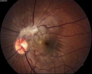
- Fundus: Abnormality as seen on picture.
What is the diagnosis? Any differential diagnoses?
Click for answer
Left combined hamartoma of the retina and RPE. Not many differentials with such characteristics fundus appearance with peripapillary pigmented low elevation with telangiectasia on the disc and preretinal fibrosis often involving the disc and macula. Only differentials worth considering in this age group may be optic disc granuloma such as toxocara.
What is your next investigation? What would be the distinguishing features in this investigation that would support your diagnosis?
Click for answer
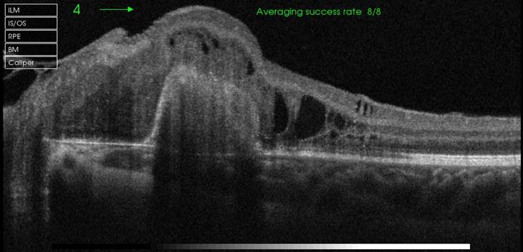
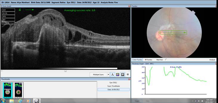
OCT with either radial or raster cut across the lesion will show distinguish loss of internal retinal architecture with a hyperreflective homogenous appeance with RPE, retinal and preretinal involvement. Variable corrugation at macula or cystic maculopathy may be seen depending on degree of macular involvement. Retinal angiography is optional often highlighting the surface telangiectasia at the disc and some late hyperfluorescence in the lesion itself. Visual field test and autofluorescence imaging are not necessary in making the diagnosis and only provide optional information with not much prognostic value.


How would you follow it up if not referred?
Click for answer
If not referred to a retinal specialist to confirm the diagnosis, I would suggest yearly follow up with photo and OCT for a few years then once confirmed no change in degree of retinal surface traction, 5 yearly review is reasonable as this lesion is almost always benign and only tractional changes may rarely progress. Surgical treatment to relieve surface traction has only extremely limited role.