Abnormal Macular with Odd-looking Disc
Case Study:
Dr Daniel Chiu
Presentation
22 yo female university science student with presents with mild binocular reading difficulty. Recently she is under a lot of exam stress and had been using steroid cream for facial rash.
Clinical findings

- VAR s 6/9 VAL s 6/5
- Anterior : Normal anterior segment and lens.
- BEO N5 but uneasy.
- IOP: Normal. No RAPD.
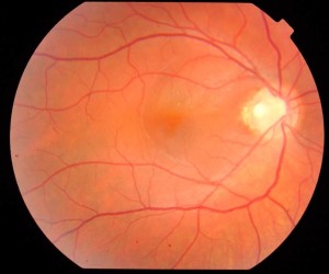
- Fundus: as per attached picture.
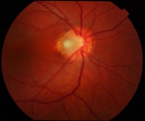
Central macular oedema or fluid noted with “odd” disc cupping according to referring optometrist
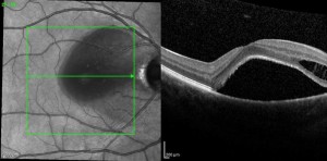
OCT scan was done.
What are the differential diagnoses? What is the correct diagnosis?
Correct diagnosis is right congenital optic disc pit with central macular detachment.
Differential diagnoses may include
- Central serous chorioretinopathy (some possible precipitating factors such as stress and steroid use but slightly young and uncommon to affect female gender)
- Vogt-Koyanagi-Harada Syndrome can also give serous detachment in the context of concomitant bilateral posterior uveitis.
What are the distinguishing features to support the correct diagnosis?
Clinical disc examination clinch the diagnosis (see enlarged disc photo). 
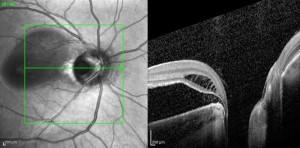
OCT confirms central detachment with characteristic relatively healthy retinal layer but detachment extends towards disc and often see mid-retinal and outer retinal schitic changes also extending to the disc.


Horizontal raster scan across the disc show cut section of disc pit really well (in this case the pit is particularly larger than usual)
Why is she 6/9 unaided but have some trouble reading?
Wtth the outer retinal layer including photoreceptor layer outer segment still quite healthy she can resolve 6/6 with a hyperopic shift just like cases of CSR. She is likely to have micropsia and imbalance in her binocular accommodation would lead to binocular reading difficulties.
Optic disc pit maculopathy or macular detachment can be very chronic in some patient and may even fluctuate in severity. Generally surgical attention is required semi-urgently.